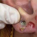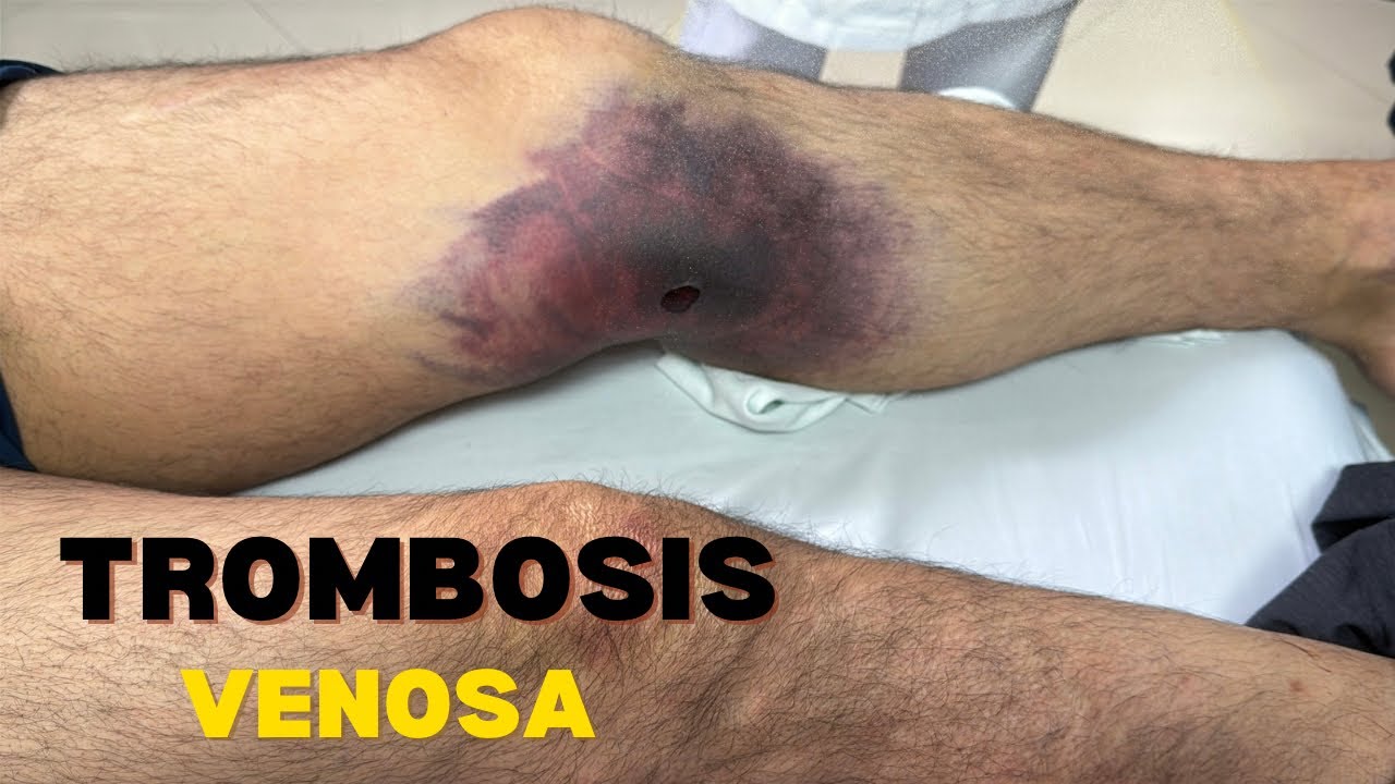
Venous Thrombosis
Please scroll down to watch the video.👇👇
✅ How to Manage and Treat Venous Thrombosis
Venous thrombosis is the formation of a blood clot in a vein, commonly in the legs (deep vein thrombosis – DVT). If untreated, the clot can travel to the lungs and cause a pulmonary embolism (PE), which is life-threatening.
⚠️ 1. Recognize the Signs and Symptoms
-
Swelling in the leg (usually one-sided)
-
Pain or tenderness, especially in the calf
-
Redness or warmth over the affected area
-
Visible veins or skin discoloration
If you suspect venous thrombosis, seek immediate medical attention.
🧪 2. Get Diagnosed
Your healthcare provider may order:
-
Doppler ultrasound (most common)
-
D-dimer blood test (helps detect clotting activity)
-
CT pulmonary angiogram (if PE is suspected)
-
Venography (less common, but very accurate)
💊 3. Begin Treatment
Treatment depends on clot location, severity, and risk of embolism.
🔹 Anticoagulants (Blood Thinners)
-
Initial: Heparin (IV or injection)
-
Long-term: Warfarin, or Direct Oral Anticoagulants (DOACs) like:
-
Apixaban (Eliquis)
-
Rivaroxaban (Xarelto)
-
Dabigatran (Pradaxa)
-
Edoxaban (Savaysa)
-
🔹 Thrombolytics (Clot Busters)
Used in severe or life-threatening cases, such as a massive PE.
🔹 IVC Filter
Inserted into the inferior vena cava to catch clots if anticoagulants can’t be used.
🧦 4. Prevent Complications
-
Compression stockings: Help reduce swelling and prevent post-thrombotic syndrome.
-
Leg elevation: Helps reduce swelling.
-
Stay active: Move your legs regularly if you’re sitting for long periods.
🛡️ 5. Prevention (If You’re at Risk)
-
Walk soon after surgery
-
Drink water during long flights or trips
-
Take prescribed blood thinners if recommended
-
Avoid smoking
-
Manage weight
📚 References
-
Kearon C, et al. Chest Guidelines for VTE Treatment, 2016.
-
American Heart Association:
-
CDC: Venous Thromboembolism
Certainly! Here’s a detailed, referenced explanation on Venous Thrombosis, including pathophysiology, risk factors, diagnosis, treatment, and scientific sources.
🩸 Venous Thrombosis: A Detailed Clinical Guide
🔍 What Is Venous Thrombosis?
Venous thrombosis refers to the formation of a blood clot (thrombus) within a vein. The most clinically significant form is Deep Vein Thrombosis (DVT), which typically occurs in the deep veins of the legs. It can lead to Pulmonary Embolism (PE) if the clot dislodges and travels to the lungs.
🧬 Pathophysiology
Venous thrombosis generally occurs due to Virchow’s Triad:
-
Venous stasis – slow or stagnant blood flow
-
Endothelial injury – damage to the blood vessel lining
-
Hypercoagulability – increased tendency of the blood to clot
These three factors disturb the balance between pro-coagulant and anti-coagulant forces in the body.
Source: Esmon CT. Basic mechanisms and pathogenesis of venous thrombosis. Blood Rev. 2009.
⚠️ Risk Factors
Type Examples Acquired Surgery, trauma, cancer, pregnancy, immobility, obesity Genetic Factor V Leiden, Prothrombin mutation, Protein C/S deficiency Lifestyle Smoking, long travel, dehydration Medications Oral contraceptives, hormone replacement therapy Source: Heit JA. Epidemiology of venous thromboembolism. Nat Rev Cardiol. 2015.
🧪 Diagnosis
-
Clinical Evaluation: Using Wells Score for DVT probability
-
D-dimer Test: Sensitive but not specific. Best used to rule out DVT in low-risk cases.
-
Duplex Ultrasound: First-line imaging for DVT.
-
CT Pulmonary Angiography: Gold standard for diagnosing PE.
-
MRI/Venography: Occasionally used for pelvic or upper extremity DVT.
Source: Kearon C, Akl EA, et al. Chest guideline and expert panel report. Chest. 2016.
💊 Treatment
1. Anticoagulation (Mainstay of Therapy)
Drug Type Examples Notes Heparin UFH, LMWH (e.g., enoxaparin) Used initially, especially in hospitals Vitamin K Antagonists Warfarin Requires INR monitoring Direct Oral Anticoagulants (DOACs) Apixaban, Rivaroxaban, Dabigatran No routine lab monitoring 2. Thrombolytic Therapy
-
Reserved for massive PE or phlegmasia cerulea dolens (severe limb-threatening DVT)
-
Examples: Alteplase (tPA)
3. Inferior Vena Cava (IVC) Filter
-
Used if anticoagulation is contraindicated
-
May prevent PE but has long-term risks (e.g., thrombosis at the filter site)
4. Compression Therapy
-
Graduated compression stockings help reduce swelling and prevent post-thrombotic syndrome
Source: Schulman S, Kearon C. Chest. 2016.
📆 Duration of Treatment
Situation Treatment Duration First unprovoked DVT/PE At least 3–6 months Recurrent VTE or thrombophilia Indefinite (lifelong) Provoked (e.g., surgery) 3 months minimum
🛡️ Prevention
-
Early mobilization after surgery or injury
-
Prophylactic anticoagulation in hospital patients
-
Compression stockings on long flights or bedrest
-
Hydration and leg movement during travel
Source: NICE Guidelines [NG158] (2020)
📚 Key References
-
Kearon C, Akl EA, et al. Antithrombotic Therapy for VTE Disease: CHEST Guideline and Expert Panel Report. Chest. 2016.
-
Heit JA. Epidemiology of venous thromboembolism. Nat Rev Cardiol. 2015.
-
Esmon CT. Basic mechanisms and pathogenesis of venous thrombosis. Blood Rev. 2009.
-
NICE Clinical Guideline NG158 (2020):
-
CDC – Deep Vein Thrombosis:
-