Mucocele (also known as an oral mucous cyst or “snot bubble”) is a fluid-filled swelling that appears on the inside of the lips, usually the lower lip. It’s caused by blocked or ruptured salivary glands, leading to mucus accumulation under the surface of the mucosa.
Here’s a detailed look at how mucocele removal is done—both medically and what not to do at home.
Reasoned about mucocele removal for a couple of seconds
Below is an in-depth explanation of mucocele (commonly known as a “snot bubble”) removal on the bottom lip, including the underlying causes, treatment options, procedural details, post-operative care, and references from reputable sources.
1. Understanding Mucoceles
What Is a Mucocele?
Definition: A mucocele is a benign, mucus-filled cyst that forms when a salivary gland duct becomes blocked or ruptured.
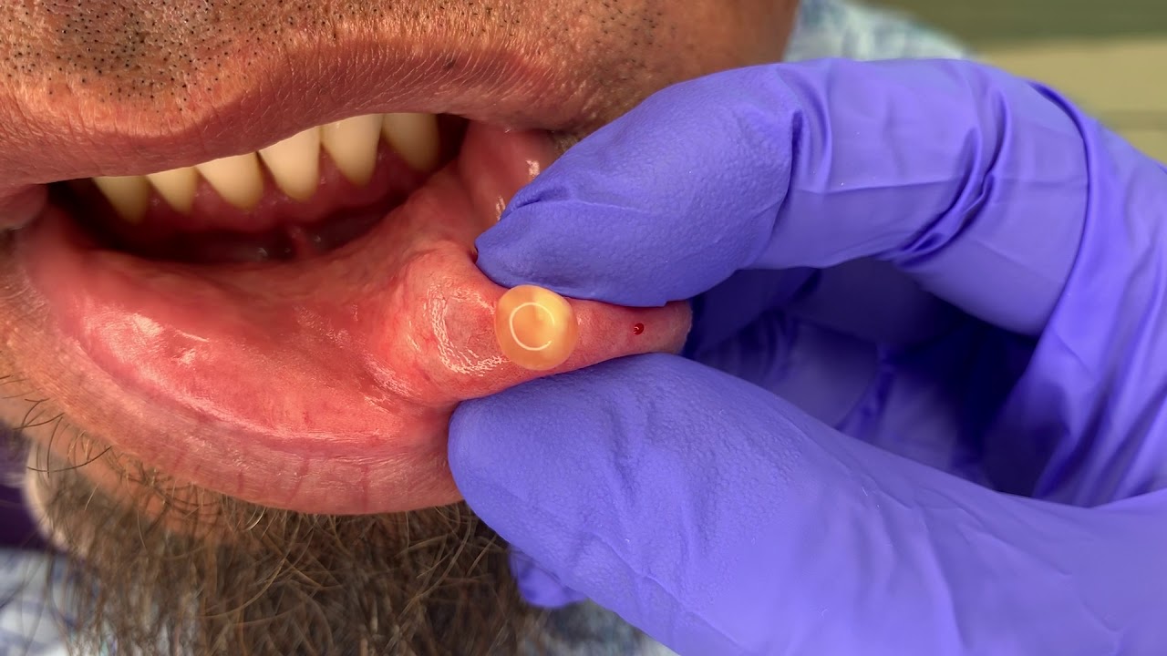
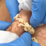
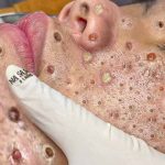
Appearance: They typically appear as a bluish, translucent, or dome-shaped lump on the inner surface of the lower lip.
Causes:
Trauma (e.g., habitual lip biting or accidental injury) leading to ductal rupture.
Blockage of the salivary gland duct that causes mucus to pool beneath the surface.
Reference: Cleveland Clinic explains that mucoceles are usually related to mechanical trauma or ductal obstruction, resulting in an accumulation of mucus under the mucosal tissue.
2. Medical Treatment Options
While some small mucoceles may resolve on their own, larger or persistent lesions are treated using one of the following methods:
A. Surgical Excision (Preferred Method)
Pre-procedure:
The area is examined, and if necessary, a diagnostic biopsy may be performed to confirm the mucocele diagnosis.
Local anesthesia: Applied to numb the region.
Procedure:
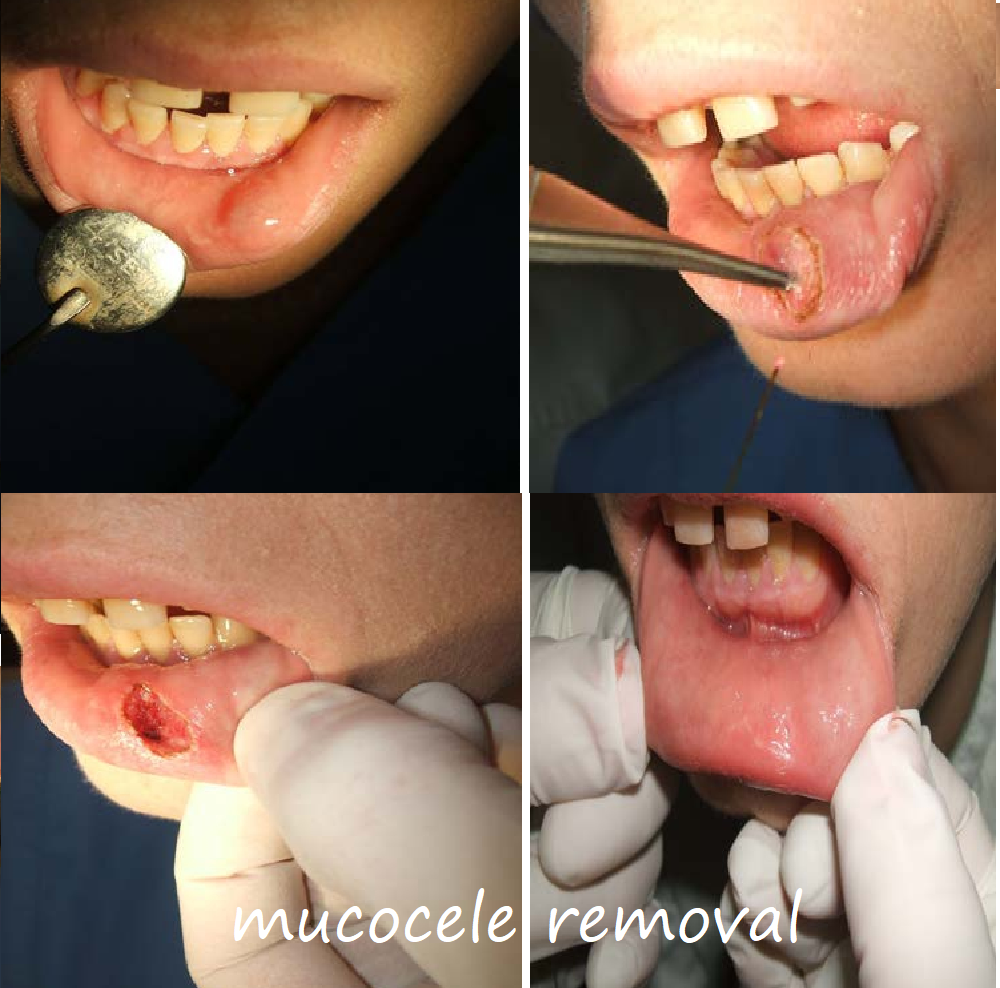
A small incision is made over the cyst.
The entire cyst is removed along with a small portion of the adjoining salivary gland tissue. This is critical because incomplete removal can lead to recurrence.
The incision may be closed with fine sutures.
Post-op care:
Patients typically experience mild discomfort or swelling.
Soft, non-irritating foods are recommended for the first few days.
Follow-up appointments are necessary to monitor healing and ensure no recurrence.
Reference: The American Academy of Oral Medicine outlines that complete removal of both the cyst and the associated gland tissue is essential to prevent future recurrence.
B. Laser Therapy
Mechanism:
A laser (often a CO₂ or diode laser) is used to vaporize the cyst.
Advantages:
Reduced bleeding due to the laser’s cauterizing effect.
Minimal post-operative discomfort and generally faster recovery.
Considerations:
Not all practitioners may have access to the necessary technology, and cost can be a factor.
C. Cryotherapy
Procedure:
Liquid nitrogen is applied to freeze the cyst.
Over subsequent sessions, the cyst is gradually reduced in size.
Usage:
More common for superficial lesions.
The risk of adjacent tissue damage necessitates precise application.
Additional details regarding laser and cryotherapy protocols can be found in clinical articles and case studies available through PubMed, which report these as viable alternatives especially when surgical excision might pose cosmetic concerns.
3. Detailed Procedural Insights
Surgical Excision Steps in Detail:
Pre-Operative Assessment:
A thorough intraoral examination is done to determine the exact size and location of the cyst.
In some cases, imaging or histopathology is used to rule out other types of lesions.
Anesthesia and Preparation:
Local anesthesia is typically sufficient for lesions on the lower lip.
The area is disinfected, and sterile draping is applied.
Incision and Excision:
The surgeon makes a precise incision over the cyst.
Using delicate instruments, the cyst is dissected from the surrounding tissues.
The surgeon ensures removal of the cyst wall and any associated glandular tissue.
Closure and Aftercare:
If the wound is small, it may heal by secondary intention; however, many lesions are sutured.
Instructions include gentle oral hygiene practices, possible antibiotic prophylaxis (if there is a risk of infection), and analgesics for pain control.
Follow-Up:
A follow-up visit is scheduled to remove sutures (if placed) and to confirm that the area is healing properly without signs of recurrence.
Reference: A study published on the NIH website details the procedure, complications, and recurrence rates, offering clinicians guidance on the best practices for surgical excision of mucoceles.
4. Post-Operative Care and Management
Dietary Recommendations:
Soft foods and avoiding spicy or acidic foods that may irritate the surgical site.
Oral Hygiene:
Gentle rinsing with saline or an antiseptic mouthwash to prevent infection.
Activity:
Avoid strenuous activity or any behavior that might disrupt the healing process (e.g., picking at the site).
Recognizing Complications:
Signs of infection (increased pain, redness, swelling, or pus formation) should prompt immediate consultation with a healthcare provider.
Reference: Post-operative care guidelines are well-documented by both the Cleveland Clinic and the American Academy of Oral Medicine, ensuring patient safety and optimal healing.
5. Conclusion
Mucocele removal on the bottom lip can be performed effectively with minimal complications when carried out by trained professionals. The key to preventing recurrence is a complete excision of both the cyst and the related salivary gland tissue. Alternative treatments like laser therapy and cryotherapy offer additional options, each with their specific benefits and considerations.
For individuals experiencing mucoceles or those concerned about a recurrent lesion, consulting with a dentist or oral surgeon is essential for a tailored treatment plan.
If you need further details on the procedure, post-operative care, or have additional questions, feel free to ask!